Your DVT Ultrasound Scan: What to Expect in the UK
You may be reading this because one leg suddenly looks different. It may feel tighter in the calf, more swollen around the ankle, warmer than usual, or more painful when you walk. That kind of change can be unsettling, especially if it seems to come from nowhere.
A dvt ultrasound scan is the test doctors use most often to check whether a blood clot is sitting in the deep veins of the leg. If you’re trying to work out whether your symptoms need urgent attention, it helps to know what this scan does, what it can and can’t show, and how it fits with earlier steps such as symptom checking and D-dimer testing.
Understanding Leg Pain and When to Suspect DVT
One-sided leg swelling, new calf pain, redness, or unusual warmth can happen for many reasons. A strained muscle, a Baker’s cyst, skin infection, or fluid retention can all cause similar symptoms. But one cause needs prompt medical attention: deep vein thrombosis, or DVT.

In plain terms, DVT means a blood clot has formed in a deep vein, usually in the leg. In the UK, DVT affects approximately 1 in 1,000 people annually, according to NCBI Bookshelf guidance on deep vein thrombosis. That makes it important enough to take seriously, but also common enough that hospitals and clinics have a very clear way to assess it.
Symptoms that deserve attention
You should think about DVT if you have symptoms such as:
- One leg swelling more than the other. Shoes, socks, or trouser legs may suddenly feel tighter on one side.
- Calf or thigh pain. People often describe it as cramping, soreness, or a heavy ache that doesn’t behave like a normal pulled muscle.
- Warmth or redness. The skin can look flushed or feel hotter over the painful area.
- Symptoms after a trigger. A long period of immobility, recent surgery, illness, or recovery time in bed can raise concern.
Why ultrasound matters
A lot of people worry that diagnosis will be complicated or invasive. Usually, it isn’t. The dvt ultrasound scan is the main test because it can look directly at the vein and help the clinician decide whether a clot is present.
Why this matters for you: the scan is designed to answer a practical question quickly. Is there a clot that needs treatment, or is another cause of leg pain more likely?
Ultrasound is especially useful for clots in the larger upper leg veins. The same NCBI source reports a diagnostic odds ratio of 379 for proximal vein DVT ultrasound, meaning a positive scan is far more likely in people who have a clot than in those who don’t. That kind of performance is why ultrasound has become the standard route to a clear diagnosis.
What Is a DVT Ultrasound Scan
A dvt ultrasound scan is a non-invasive imaging test that uses sound waves to look inside your leg veins. There are no needles going into the vein for the scan itself, and there is no radiation. If you’ve had a pregnancy scan or an abdominal ultrasound before, the basic idea is similar, but the focus here is blood vessels rather than organs.
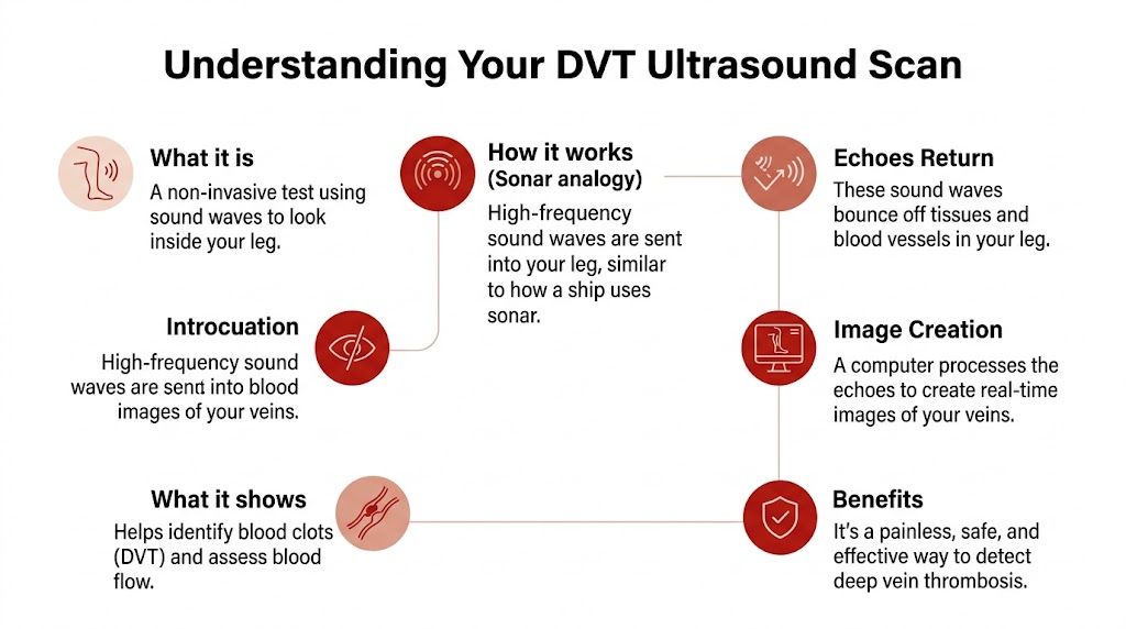
A simple way to picture it
The easiest analogy is sonar. A ship sends out sound, the sound bounces back, and the returning echoes help map what’s in the water. Ultrasound works in much the same way. The probe sends high-frequency sound waves into your leg, and a computer turns the returning echoes into a live image.
That live image lets the clinician examine the shape of the vein and look for signs that blood isn’t flowing normally.
The two parts patients often hear about
People sometimes hear several terms during the appointment and aren’t sure whether they mean different tests. Usually, they’re describing parts of the same examination.
Compression ultrasound
This is the key practical check. A healthy vein should gently flatten when the sonographer presses with the probe. If part of the vein doesn’t compress, that raises concern that a clot is occupying the space.
Think of an empty soft straw versus one packed with material. The empty one gives way. The blocked one doesn’t.
Duplex ultrasound
Duplex ultrasound adds flow assessment to the picture. It combines the structural view with Doppler techniques, which help the operator assess how blood is moving through the vein. That can help show whether flow is reduced, altered, or interrupted.
This matters because a clot isn’t only a lump in a vessel. It also changes how blood travels through that vessel.
Why it is the first-line test
Doctors favour ultrasound because it answers the main question without exposing you to a more invasive test. It can be done in hospital imaging, emergency care, and in some settings at the bedside by trained clinicians.
For a patient, the practical advantages are straightforward:
- It doesn’t involve surgery
- It doesn’t use radiation
- It can be done relatively quickly
- It gives information that directly guides treatment decisions
A dvt ultrasound scan is less about “taking a picture” and more about checking whether the vein behaves like a normal vein should.
That’s often where confusion clears. The operator isn’t just looking for something obvious on a screen. They’re actively testing the vein, segment by segment, to see whether it compresses and whether blood flow looks right.
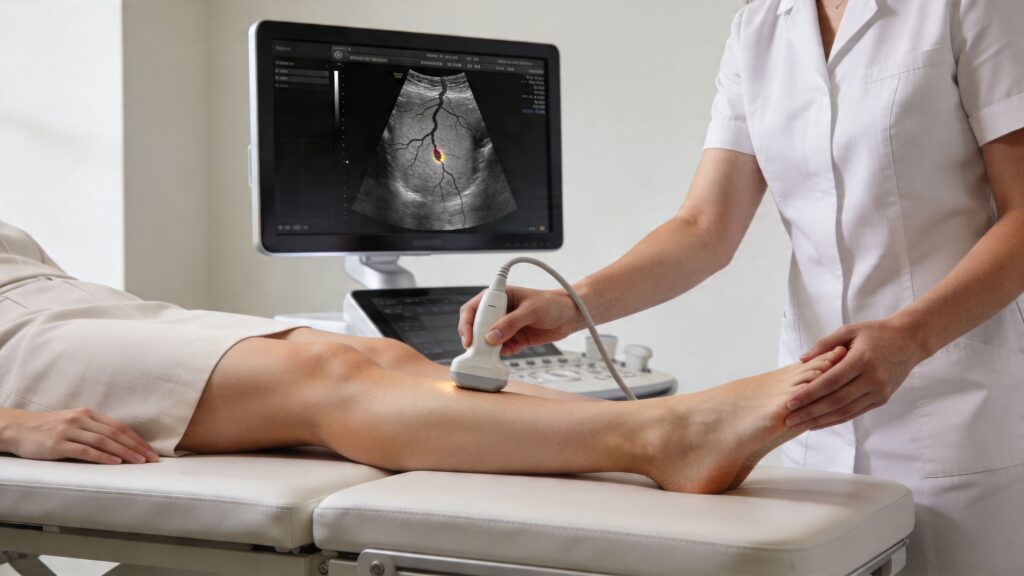
How the DVT Ultrasound Scan Is Performed
The day of the scan is usually much simpler than people expect. You don’t normally need to fast, and in most cases you can eat, drink, and take your usual medicines unless the clinic has told you otherwise for a separate reason.

Before the scan starts
You’ll usually be asked to uncover the affected leg and lie on an examination couch. Loose clothing helps. If you’ve come straight from work in tight trousers or shapewear, the appointment can feel more awkward than it needs to.
The room often looks fairly ordinary: couch, screen, ultrasound machine, gel, and towels or paper roll. It isn’t an operating theatre. That’s reassuring for many people.
What the sonographer actually does
The standard procedure is a complete duplex ultrasound with compression at 2-cm intervals from the inguinal ligament to the ankle, using high-frequency linear transducers in the 7.5-12 MHz range, as described in CSV guidance on lower limb venous duplex assessment.
That sounds technical, but the patient experience is simple. The sonographer puts gel on the skin, places the probe over the leg, and moves steadily along the vein pathway. At regular points, they press down firmly to see whether the vein closes properly.
What you may feel
Individuals often notice three things:
- Cool gel on the skin. It can be a bit chilly at first.
- Pressure from the probe. This is often the part patients mistake for “pain from the scan”, but it’s really the pressure needed to test compressibility.
- Frequent pauses. The sonographer may stop, adjust the angle, or check the same area twice.
If your leg is already sore, firm pressure can be uncomfortable. But the scan itself isn’t damaging the vein or moving a clot around.
Practical rule: if the pressure feels too much, say so. The sonographer still needs to assess the vein, but they can explain what they’re doing and work as gently as possible.
Why the scan covers more than the painful spot
People often expect the scan to focus only on the reddest or most painful area. In reality, the operator follows the deep veins in an organised path. That’s because the clot may sit above or below where you feel symptoms.
For example, someone may point to pain behind the knee, but the important finding may be higher up in the thigh. That is also why the examination can seem methodical rather than symptom-led.
A useful comparison
If you’ve ever read about another screening ultrasound, such as abdominal aortic aneurysm screening, the logic is similar. The clinician follows a set anatomical route instead of scanning only where you feel discomfort. That approach improves the chances of finding what matters.
After the images are taken
Once the sonographer has enough information, the gel is wiped away and you can usually get dressed straight away. Some patients receive preliminary information immediately. In other settings, the images are reviewed and the result is then passed to the doctor responsible for your care.
The key point is that the test is built to answer a practical clinical question quickly. For most patients, the hardest part is the anxiety beforehand, not the scan itself.
Interpreting Your Scan Results and Limitations
When the scan is finished, the result usually falls into one of three groups: positive, negative, or inconclusive. The wording may vary, but the meaning is similar.
If the scan is positive
A positive result means the scan has found signs of a clot in the deep veins. The next step is usually urgent clinical review and treatment planning. In many cases that means anticoagulant medication, often called blood thinners.
This result answers the question clearly. The focus then shifts from diagnosis to reducing the chance of the clot growing or travelling.
If the scan is negative
A negative result means no DVT was seen on that examination. That can be very reassuring, but it doesn’t always mean the story ends there.
A negative scan may point the clinician toward other causes of symptoms, such as muscle injury, superficial vein problems, joint issues, or infection. It may also be combined with blood tests and your symptom history to decide whether further follow-up is needed.
If the scan is inconclusive
Sometimes the view isn’t clear enough to rule a clot in or out with confidence. That may happen because of body shape, swelling, tenderness, or the exact location of concern.
In that situation, the next step may be a repeat ultrasound, another test, or review by a specialist team.
If your symptoms are convincing but the result feels uncertain, ask what the plan is next. “What happens if the pain or swelling continues?” is a sensible question.
Where ultrasound is strongest, and where caution matters
UK-specific data show ultrasound is highly accurate for proximal, or thigh, veins with 98% specificity, but sensitivity for distal, or calf, DVT can drop to 43%, which is why a repeat scan in 5-7 days may be needed if suspicion remains high, according to this review in PMC.
That sounds technical, so its real-life meaning is:
- Large clots in bigger upper-leg veins are easier to confirm
- Small calf-vein clots can be harder to detect
- A single negative scan doesn’t always close the case if symptoms and risk factors still point toward DVT
Questions worth asking after your result
- Was the scan complete or limited?
- Do I need a repeat scan if symptoms continue?
- Should I have blood tests as well?
- What else could be causing these symptoms?
If you’re arranging blood work privately or reviewing earlier results, services that offer online lab tests in the UK can help you gather supporting information, but they don’t replace formal imaging when DVT is suspected.
The Role of At-Home Testing and Other Diagnostics
Ultrasound is the formal diagnostic test, but it isn’t always the first step in practice. Many people start at home by noticing symptoms, checking whether one leg looks different, and asking whether they need urgent medical assessment.
That earlier stage matters. It can help you decide whether you likely need emergency care, same-day review, or a more measured next step such as a blood test.
Where D-dimer fits in
A D-dimer test looks for substances released when the body forms and breaks down clots. It doesn’t tell you exactly where a clot is, and it can’t confirm DVT on its own. What it can do well is help rule DVT out in the right clinical context.
A clinical study summary reported that diagnostic algorithms using D-dimer thresholds can safely rule out DVT and “reduce the need for ultrasound imaging by nearly 50%”, as described in this report on AI-guided ultrasound and DVT pathways.
For patients, that’s useful because it creates a clearer path:
- Low concern plus reassuring D-dimer may mean no urgent scan is needed
- Higher concern or a positive D-dimer usually pushes the decision toward ultrasound
- Ongoing symptoms despite an earlier low-risk view still deserve review
Comparing DVT diagnostic methods
| Method | What it Measures | Best For | Pros | Cons |
|---|---|---|---|---|
| Symptom and risk assessment | Your symptoms, history, and clinical risk | First decision-making step | Quick, helps guide what comes next | Can’t confirm or exclude DVT by itself |
| D-dimer blood test | Blood markers linked to clot formation and breakdown | Ruling out DVT in appropriate patients | Can reduce unnecessary imaging | Doesn’t show where a clot is and doesn’t confirm DVT alone |
| DVT ultrasound scan | Vein compressibility and blood flow | Confirming or excluding suspected leg DVT | Non-invasive and directly useful for treatment decisions | Can be less sensitive for some calf clots |
| Venography | Contrast imaging of the veins | Selected complex cases | Detailed vein imaging | Invasive, so it’s not the usual first choice now |
The bridge between home and clinic
Many readers want a practical answer to one question: What can I do before I reach hospital imaging? The answer is to use home information wisely, not to self-diagnose.
That means noticing patterns such as one-sided swelling, timing after surgery or immobility, and whether symptoms are worsening. If a clinician thinks blood testing is appropriate, broader private health screening in the UK may form part of your overall health picture, but suspected DVT still needs a proper clinical pathway.
The safest mindset is this. Home steps can help you decide how urgently to seek care. They do not replace the scan when a clot is genuinely possible.
Emergency Signs of Pulmonary Embolism
A clot in the leg becomes far more dangerous if part of it travels to the lungs. That complication is called a pulmonary embolism, or PE.

This is the point where you shouldn’t monitor symptoms at home or wait for a routine appointment. Trained UK emergency physicians using point-of-care ultrasound achieve pooled sensitivity of 96% for DVT, and the reason speed matters is that pulmonary embolism can occur in up to 50% of untreated proximal DVT cases, as noted in the ASRA discussion of lower extremity DVT scanning.
Call 999 or go to A&E now if you have
- Sudden shortness of breath that comes on without a clear explanation
- Sharp chest pain, especially if it gets worse when breathing in
- Dizziness or feeling faint
- Coughing up blood
- A fast deterioration after leg swelling or calf pain
Don’t wait to see if it settles
If leg symptoms are followed by breathing symptoms, chest pain, or collapse, treat that as an emergency. The question is no longer whether you might need a dvt ultrasound scan soon. The question is whether a clot may already have moved.
New chest symptoms with suspected DVT need urgent emergency care, not home observation.
Your DVT Scan Questions Answered
Is a dvt ultrasound scan painful
Usually no. The gel is cool, and the probe pressure can feel uncomfortable if your leg is already tender, but it is generally well-tolerated.
Do I need to fast
In most cases, no. You can usually eat and drink normally unless you’ve been given different instructions for another test.
Can I drive afterwards
Usually yes, because the scan itself doesn’t involve sedation. If your leg is very painful or you’ve been told you may need immediate treatment, it may still be wise to arrange help.
How long does it take
It varies by clinic and by how much detail the operator needs, but patients are often surprised by how straightforward it is.
If my scan is negative but I still feel unwell, what then
Go back for review, especially if swelling or pain persists or worsens. If you also develop breathlessness, chest pain, or you’re unsure whether another condition could be involved, this guide on what causes shortness of breath is a useful general resource, but urgent symptoms still need direct medical assessment.
Can a blood test replace the scan
Not by itself. A D-dimer can help with triage, but ultrasound is the test that looks directly at the vein when DVT is a real possibility.
If you want a convenient starting point for home-based health checks, Repose Healthcare offers private at-home testing across the UK and Republic of Ireland, with clear instructions, UK-accredited laboratory processing, and secure online results. If you’re weighing symptoms, monitoring your health, or looking for a practical first step before speaking to a clinician, their testing options can help you make a more informed decision about what to do next.
You Might Also Like
The 2026 Cost of Hepatitis Vaccination in the UK
You're usually looking up the cost of hepatitis vaccination for a practical reason. A trip [...]
Hormone Blood Test London: Find Your Perfect Test
Some London health questions start subtly. Your cycle changes a little. You feel wired at [...]
Peanut Allergy Test Kit: A Practical UK User’s Guide
A peanut reaction often starts with uncertainty, not clarity. You eat a biscuit, satay sauce, [...]
NIPT Test Edinburgh: Your 2026 Guide
If you're searching nipt test edinburgh, you're probably already in that familiar stage of pregnancy [...]
Peripheral Artery Disease UK: Peripheral Artery Disease UK:
You might be reading this because walking has started to feel different. Perhaps your calf [...]
NIPT Test Cardiff: Clinic, Home & NHS Pathways 2026
If you're in Cardiff, newly pregnant, and staring at screening options that all seem to [...]
Gender Scan Difference UK: Accuracy, Timing & Cost Compared
You've probably already done the same search most parents do. One tab shows the NHS [...]
NIPT Test Cambridge: Guide to Options & Booking 2026
You're in Cambridge, you've had the first rush of finding out you're pregnant, and then [...]
Home COVID Test Results: A Practical UK & Ireland Guide
You've done the swab, mixed the sample, added the drops, and now you're staring at [...]
DVT Test Squeeze Calf: Homan’s Sign & Beyond
You notice one calf is sore, a little swollen, and not behaving like an ordinary [...]
Your Guide to Every Travel Clinic Leicester Offers in 2026
Flights are booked. The hotel is sorted. Your passport is still in date. Then one [...]
13 Week Gender Scan: Accuracy, NIPT & What to Expect
By the time you reach 13 weeks, pregnancy often starts to feel more real in [...]
Your Pregnancy Scan London: Guide to Options & Costs
A positive test can make London feel suddenly very small and very complicated. You might [...]
Your Nuchal Dating Scan Explained: A 2026 UK Guide
That appointment message has landed in your phone or come through the post, and suddenly [...]
Normal Cortisol Levels UK: Get Clarity
Some people search for normal cortisol levels uk after weeks of feeling unlike themselves. They’re [...]
How to Test Iron Levels in Blood: A Complete UK Guide
You’re probably not searching how to test iron levels in blood out of curiosity. More [...]
Growth Scan Pregnancy: UK Guide & What to Expect
You’re at an antenatal appointment, everything seems routine, and then your midwife says, “We’d like [...]
Gender Scan Near Me: Your 2026 Guide to Options
You’re probably here because the question has stopped feeling abstract. Maybe you’ve had your booking [...]
Progesterone Test For Pregnancy: Viability & Home Kit
The positive test is there, but relief doesn’t always follow. For many women, early pregnancy [...]
Ovulation Blood Tests: Confirm Your Ovulation
You’ve probably already done some version of this. You’ve checked an app, watched for a [...]
How to Test Testosterone Levels: A UK Guide
You wake up tired, despite a full night in bed. Your training feels flat. Your [...]
UK Vitamin D Test: A Complete At-Home Guide for 2026
You wake up tired, even after a full night’s sleep. By mid-afternoon you’re reaching for [...]
Blood Iron Level Test: A Complete UK Guide for 2026
Some days it isn’t dramatic. You’re just slower than usual. You wake up after a [...]
Cortisol Levels Test: Your Comprehensive UK Guide
You wake up tired, push through the morning on caffeine, feel oddly tense by midday, [...]
Travel Clinic Leeds: Compare 7 Top Options For 2026
You book the flights first. Then the practical questions start. Is a tetanus booster enough [...]
Private Blood Test Leicester: A Complete 2026 Guide
You wake up tired again. Maybe you’ve had weeks of bloating, odd fatigue, headaches, low [...]
Genetic Testing Before Pregnancy: A UK Guide for 2026
You might be at the stage where baby plans still live in half-finished conversations. One [...]
Harmony NIPT Test: A Clear Guide for Expectant Parents
You may be sitting with a booking slip from your midwife, a browser full of [...]
Decode Your Health: Understanding Full Blood Count Results
That moment is familiar. You open your blood test report on your phone, see a [...]
Iron Level Tester: Your Guide to Better Health
You wake up tired, push through the day with coffee, and still feel flat by [...]