7 Week Scan Pictures: See Early Development
You get a positive test at home, then wait days for a scan appointment, and those two parts of early pregnancy can feel like they belong to different worlds. At home, you are reading hormone changes. In the clinic, you are looking for structures on a screen. A 7 week scan helps connect those pieces.
At this stage, home testing and ultrasound are answering different questions. Biochemical tests such as hCG and progesterone can show that pregnancy hormones are rising, and some people also use a progesterone test for pregnancy to follow that early hormonal picture. Ultrasound adds the visual side. It shows whether there is a pregnancy sac in the uterus and whether early features such as the yolk sac, fetal pole, and cardiac activity are visible for this point in pregnancy.
A simple way to think about it is that home tests are like early signals, while the scan is the first map. One tells you the body is producing pregnancy hormones. The other shows where the pregnancy is developing and how the pregnancy is measuring against your dates. That matters if your cycle length varies, you ovulated later than expected, or your test results have left you with more questions than reassurance.
In UK care, a 7-week scan is usually an early reassurance or problem-solving scan rather than the routine dating scan, which is commonly done closer to 12 weeks. The routine anomaly scan is generally offered later in pregnancy, at around 18 to 21 weeks, according to the NHS overview of ultrasound scans in pregnancy. That helps explain why a 7-week appointment can feel so important when it is arranged for bleeding, pain, fertility treatment, uncertain dates, or a previous miscarriage.
If your partner is trying to understand the milestones too, Hera Fertility resources for expectant fathers can help put the journey into context.
1. Gestational Sac Measurement at 7 Weeks
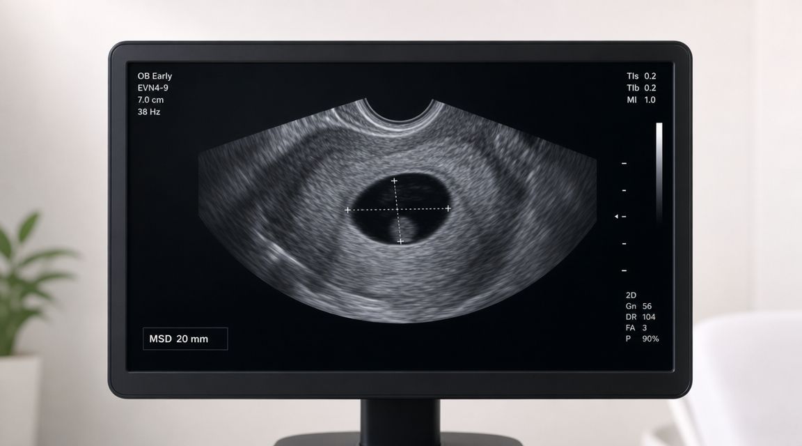
You may arrive for a scan after several days of watching home pregnancy tests darken and still feel unsure what, exactly, the sonographer is looking for. One of the first landmarks we check is the gestational sac. It is the early fluid-filled space where the pregnancy is developing, and on ultrasound it usually appears as a dark round or oval area within the uterus, outlined by a brighter rim.
By 7 weeks, the sac should usually be clearly visible on a transvaginal scan, and clinicians often record its mean sac diameter as part of the early assessment, as described in the StatPearls overview of sonography in first-trimester bleeding. The exact number matters less than the full picture. We do not read the sac size in isolation. We compare it with your dates, the appearance of other early structures, your symptoms, and what your home testing has already suggested.
What this means on your picture
For many women, this is the first point where a biochemical result becomes a visible pregnancy. Home tests show that your body is producing pregnancy-related hormones. The scan adds location and structure. It confirms that the pregnancy sac is sitting in the uterus and gives the team an early reference point for how the pregnancy is measuring.
A simple comparison helps here. Hormone tests are like seeing smoke and knowing a fire exists somewhere. Ultrasound is the part that shows where the fire is and how it is developing. That is why a scan can answer questions that a positive test alone cannot, especially if your cycle was irregular, ovulation may have happened later, or your dates feel uncertain.
If the gestational sac measures smaller than expected, your clinician may consider later ovulation before assuming there is a problem. If it measures larger, the pregnancy may be a little further along than first thought. Small differences are common in early pregnancy because a few days can change what is visible on the screen.
Practical rule: Home hormone testing shows that pregnancy activity is present. Ultrasound shows where the pregnancy is developing and whether the visual findings fit the timing.
If you want to compare what appears on the scan with the hormone side of the picture, an at-home progesterone test for pregnancy from Repose Healthcare can add context while you wait for follow-up care. It does not replace ultrasound, but it can help your at-home results and your clinical scan make more sense together.
2. Yolk Sac Visualisation and Size Assessment
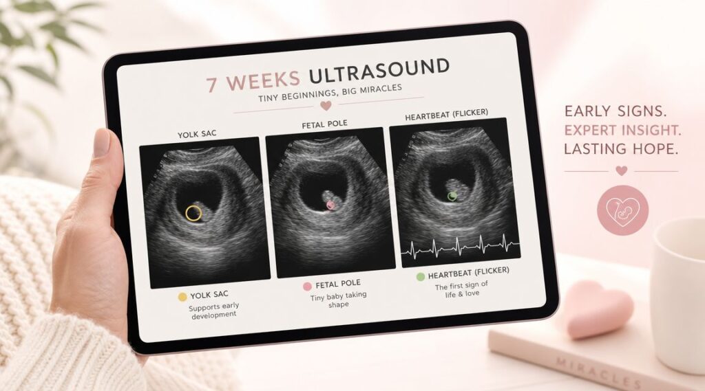
Once the sonographer identifies the gestational sac, the next important feature is often the yolk sac. In 7 week scan pictures, it usually appears as a small bright ring within the darker gestational sac.
This is one reason early scans can look very different from the polished baby images people see later in pregnancy or on social media. At this stage, the scan is less about a recognisable baby-shaped picture and more about checking for the right early structures in the right place, as explained in UK-facing guidance on why very early scans can look blurry or incomplete.

Why the yolk sac matters
The yolk sac is one of the clues that the pregnancy is developing inside the uterus and following an expected early pattern. If you had a positive test at home and were worrying because your symptoms felt mild, the presence of a yolk sac gives the scan more meaning than the picture alone.
A common real-life scenario is someone arriving anxious because their friend's scan looked “clearer.” Often, the difference is due to timing, scan angle, body position, or whether the scan was done internally. A less photogenic image doesn't automatically mean a problem.
- If you can see a small ring inside the sac: That's often the yolk sac the sonographer is checking.
- If your image looks sparse: Early scans commonly do. The sonographer is looking for key markers, not a perfect keepsake image.
- If the report mentions follow-up: That can be a normal response to uncertainty in very early pregnancy, especially when dates may be off.
If you've been tracking hCG at home, the yolk sac can feel like the bridge between what your tests suggested and what the scan can now confirm visually.
3. Embryonic Pole and Crown-Rump Length Measurement
A common 7-week moment goes like this. You have had positive tests at home, your hCG pattern suggested things were progressing, and now you are waiting to see the first structure that clearly represents the embryo itself. On the scan, that structure is the embryonic pole, also called the fetal pole. It usually appears as a tiny curved shape sitting close to the yolk sac, not as a recognisable baby outline.
By this point, the sonographer is often able to measure crown-rump length, or CRL. This is the distance from the top of the embryo to its lower end. It works like an early pregnancy ruler because it gives a practical way to estimate gestation when period dates are uncertain, cycle length varies, or conception happened later than expected.
How to read the measurement simply
CRL does not include the yolk sac, the surrounding sac, or tiny early limb buds. The sonographer is measuring the embryo itself. At 7 weeks, that measurement is still very small, which helps explain why the picture can look grainy or why an internal scan may give a clearer view.
A difference of only a day or two can change what is visible. That is why a scan can sometimes revise the timeline you built from ovulation tracking, symptoms, or home hormone tests. Your at-home testing journey and the ultrasound are not competing sources of information. They answer different questions. hCG tests show that pregnancy hormones are rising. The scan shows where the pregnancy is and how the embryo is developing visually.
When dates are unclear, CRL often gives a steadier estimate of pregnancy timing than counting from the last period alone.
If you have irregular cycles, conceived after fertility treatment, or are unsure of ovulation timing, this part of the exam often brings the clearest dating information so far. Repose Healthcare explains how early viability scans assess growth and timing in early pregnancy. If you're thinking ahead to later scan milestones, Repose Healthcare also explains the next stage through its nuchal and dating scan information.
4. Fetal Cardiac Activity and Heart Rate Documentation
For many parents, this is the point when the pregnancy stops feeling like a set of numbers on a test strip and starts looking real on a screen. After days or weeks of watching hCG results rise at home, the scan adds something those results cannot show. Motion.
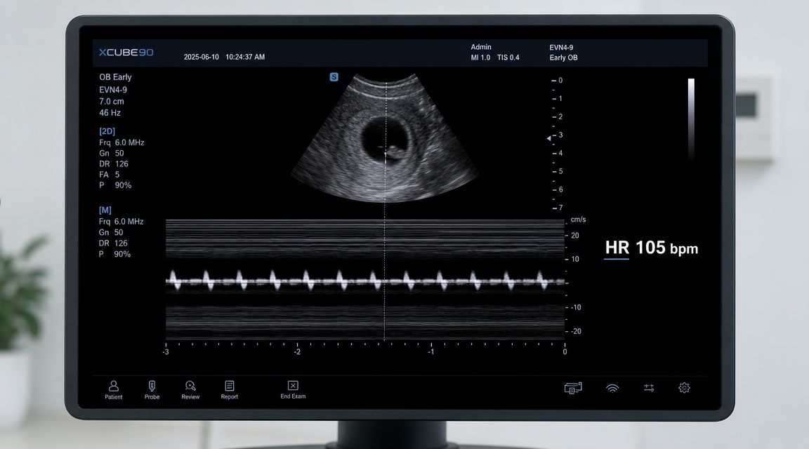
At 7 weeks, the sonographer is usually looking for a tiny flicker within the embryo. That flicker is fetal cardiac activity. It does not look like the heart shapes shown in diagrams or baby apps. On the screen, it is often a small rhythmic pulse inside the fetal pole, easy to miss if no one points it out.
A useful way to understand the difference is this. Home pregnancy testing answers the biochemical question, "Is the pregnancy hormone increasing?" Ultrasound answers the visual question, "Can we see the pregnancy developing in the uterus, and can we see cardiac activity?" Together, those two pieces give a fuller early-pregnancy picture than either one alone.
What the sonographer is documenting
The scan is not just checking whether a flicker is present. The sonographer may also document the fetal heart rate and whether the appearance matches the estimated gestational age. That matters because early pregnancy assessment is about pattern and context, not one isolated sign.
If the heart rate is measured, it is usually done by capturing the movement carefully on the live scan rather than relying on the printed image alone. A still photo can make this part seem underwhelming at home. The live examination tells far more than the printout.
Clinical takeaway: At this stage, confirmation of cardiac activity usually matters more than how clear the keepsake image looks.
This can be especially reassuring after uncertain home results, spotting, previous loss, or cycle dates that do not line up neatly. Once cardiac activity is seen at an early scan, the outlook is generally more reassuring than before that point, although the sonographer will still interpret the finding alongside growth measurements and symptoms rather than treating it as a guarantee.
If you have been referred for early reassurance, services that focus on early viability scan assessment in early pregnancy are designed to look at these findings together. The key message is simple. Your at-home tests and your 7-week ultrasound are not competing versions of the story. One shows the chemistry. The other shows the structure and the flicker of early development.
5. Amniotic Cavity Visualisation and Fluid Assessment
This is one of the less talked-about parts of 7 week scan pictures, mainly because most patients aren't shown it in a dramatic way. The sonographer sees more than the final printout suggests.
At this stage, the scan is checking the tiny internal layout of the pregnancy. That includes the relationship between the embryo and the fluid-filled spaces around it. It's often challenging to confidently identify the amniotic cavity in a photo when viewing it at home, and that's completely normal.
Why this part feels confusing
A common comment after an early scan is, “I can't tell what I'm looking at.” That's because early ultrasound is about interpretation, not obvious visual recognition. The picture may contain several shades of black, grey and white that only make sense when the sonographer explains what each area represents.
For someone who has gone from home tests to a scan, this can feel like a sudden jump in complexity. One week you're checking lines or lab values. The next, someone is discussing sacs, poles, cavities and cardiac activity.
- Dark areas often represent fluid: That's why the overall image can look hollow or empty in places.
- Tiny anatomy can overlap visually: Early structures are close together and can blur into one another on still images.
- The printed picture is only one moment: The live scan gives the sonographer much more information than the single image you take home.
This is also why two patients can leave with very different-looking 7 week scan pictures even when both pregnancies are progressing normally. The quality of the image isn't the same thing as the quality of the pregnancy.
6. Uterine Appearance and Location of Gestational Sac Positioning
One of the most important questions in early pregnancy is simple. Is the pregnancy in the uterus?
That's why the sonographer doesn't only look at the sac itself. They also assess where it sits and whether the surrounding appearance fits with an intrauterine pregnancy. In UK early-pregnancy practice, a 7-week scan is typically performed transvaginally because image resolution is better when the embryo is still very small, and viability assessment focuses on confirming an intrauterine gestational sac, yolk sac and embryonic cardiac activity, as outlined in guidance on early transvaginal scanning in the first trimester.
Why location matters as much as appearance
If you've had pain, spotting, or uncertain dates, this part of the scan can be the most medically important part even if it doesn't produce the nicest picture. A pregnancy test at home can tell you that hCG is present. It can't show whether the pregnancy is located correctly.
Real-life example. Someone may have strong positive tests and still be sent for urgent early scanning because the concern isn't whether they're pregnant. It's where the pregnancy is and whether the findings match their symptoms.
A reassuring 7-week scan isn't just about seeing something. It's about seeing the right things in the right place.
This is also why a transvaginal scan is so commonly used early on. The aim isn't to make the experience more medical than it needs to be. It's to get a clear enough view to answer the questions that matter.
7. Cervical Assessment and Adnexal Evaluation
The scan doesn't stop once the sonographer finds the embryo and heartbeat. They also look around the pregnancy.
That includes the cervix and the adnexa, meaning the ovaries and nearby structures. Patients don't always realise this because the printed image usually focuses on the pregnancy itself, but a careful early scan often includes a broader check for anything that might explain pain, bleeding, or uncertainty.
What the sonographer is checking in the background
If you're having one-sided pain, the adnexal review is especially relevant. The sonographer may look for the ovaries, note whether anything appears unusual, and build that into the clinical picture. If you've had a corpus luteum cyst mentioned before, that can also come up during early pregnancy scanning.
The cervix is usually noted with less emphasis at this stage, but it's still part of the overall assessment. In practical terms, this wider scan helps clinicians decide whether your symptoms fit with a normal early pregnancy, whether follow-up is needed, and whether the pregnancy location and surrounding anatomy make sense together.
- For bleeding or pain: Extra views outside the gestational sac can be just as important as the sac itself.
- For previous ectopic pregnancy concerns: The sonographer often pays close attention to the areas around the uterus.
- For reassurance after home testing: Clinical imaging adds information that no at-home kit can provide on its own.
If your report mentions repeat imaging, that doesn't always mean bad news. Early pregnancy often requires a second look when the first scan answers some questions but not all of them.
7-Point Comparison: 7-Week Scan Images
| Scan/Assessment | Complexity 🔄 | Resource requirements ⚡ | Expected outcomes 📊 | Ideal use cases 💡 | Key advantages ⭐ |
|---|---|---|---|---|---|
| Gestational Sac Measurement at 7 Weeks | Moderate, transvaginal skill needed | TVUS (transvaginal probe), basic measurement tools | Early intrauterine confirmation; dating ±3–5 days | Early pregnancy confirmation; fertility monitoring | Earliest sac visualisation; helps exclude ectopic |
| Yolk Sac Visualisation and Size Assessment | Moderate, small target, needs resolution | High-quality TVUS, experienced sonographer | Confirms intrauterine pregnancy; lower miscarriage likelihood when normal | Viability checks; suspected blighted ovum | Definitive intrauterine marker; simple size metric (3–6 mm) |
| Embryonic Pole & CRL Measurement | Moderate–high, precise plane & technique | TVUS with calipers, trained operator | Most accurate first-trimester dating; confirms fetal cardiac activity | Precise dating; early growth/abnormality detection | Gold‑standard dating; reproducible growth tracking |
| Fetal Cardiac Activity & Heart Rate Documentation | High, real‑time measurement, repeat verification | Real‑time ultrasound, M‑mode, skilled sonographer | Definitive viability confirmation; HR ~90–120 bpm at 7 wks | Viability confirmation after positive hormones | Definitive viability marker with prognostic value |
| Amniotic Cavity Visualisation & Fluid Assessment | Low–moderate, subjective early assessment | Standard TVUS, operator familiarity with anatomy | Confirms progressing intrauterine environment; baseline fluid status | Anatomical baseline; follow‑up of embryonic environment | Demonstrates amniotic compartment & embryonic movement |
| Uterine Appearance & Gestational Sac Positioning | Moderate, anatomical assessment, note variants | TVUS, Doppler optional, operator experience | Excludes ectopic; identifies uterine pathology affecting pregnancy | Rule out ectopic; patients with fibroids or uterine anomalies | Confirms intrauterine location; assesses maternal factors |
| Cervical Assessment & Adnexal Evaluation | Moderate, multi‑structure exam | TVUS, possible follow‑up imaging, experienced operator | Documents corpus luteum; detects adnexal complications | Correlate progesterone source; investigate adnexal symptoms | Identifies corpus luteum; excludes adnexal pathology impacting pregnancy |
Your Early Pregnancy Journey From Home Tests to Scan
A 7-week scan can feel like the first time pregnancy becomes visible. Before that, many people are working with symptoms, test lines, and maybe home hormone tracking. Once the scan happens, those clues start to line up with actual structures on the screen. You're no longer dealing only with “am I pregnant?” but with where the pregnancy is, how it's dating, and whether the expected early signs are there.
That's why 7 week scan pictures carry so much emotional weight. Even when the image looks blurry or underwhelming, the sonographer may be seeing exactly what they need to see. A gestational sac in the uterus, a yolk sac, a fetal pole, a measurable CRL, and cardiac activity can all matter more than whether the picture looks impressive on paper.
If you've been using at-home testing, it helps to think of the process in two halves. Home tests give early biochemical information. Ultrasound gives visual confirmation. Neither replaces the other. Together, they often provide a fuller picture of early pregnancy than either one alone.
It's also worth remembering that a 7-week scan is often done for a specific reason in the UK setting, not merely as a routine keepsake appointment. It may be arranged because of symptoms, uncertain dates, fertility treatment, or a previous loss. That context affects what the sonographer is prioritising. They're not just trying to capture a nice image. They're trying to answer the most important clinical questions safely and clearly.
If your scan doesn't show everything you expected, don't try to interpret the printout alone. Very early imaging has a normal range of appearances, and timing makes a big difference. If you're worried after a scan, speak with your clinician or early pregnancy service promptly.
For people who want more context while moving from home testing to clinical care, Repose Healthcare offers at-home hormone and health testing that may help support informed conversations with your clinician. And if you're also thinking about comfort during pregnancy, some readers find supportive services such as maternity massage in Edinburgh helpful later in the journey.
If you'd like to explore at-home testing alongside your clinical care, Repose Healthcare offers UK-based health tests including hormone and fertility options that may help you track early changes before and between appointments.
You Might Also Like
7 Week Scan Pictures: See Early Development
You get a positive test at home, then wait days for a scan appointment, and [...]
Home Allergy Test Kit Reviews: A UK Expert Guide 2026
You've got symptoms that don't fit neatly into one box. Maybe it's a rash after [...]
NIPT Test Sheffield: Your 2026 Guide to Options & Costs
You've had the booking confirmation for your early pregnancy appointment. You're checking dates, comparing clinics, [...]
Private Scan London: 2026 Guide to Costs & Booking
You wake up with the same worry you had last week. The pelvic pain is [...]
The 2026 Cost of Hepatitis Vaccination in the UK
You're usually looking up the cost of hepatitis vaccination for a practical reason. A trip [...]
Hormone Blood Test London: Find Your Perfect Test
Some London health questions start subtly. Your cycle changes a little. You feel wired at [...]
Peanut Allergy Test Kit: A Practical UK User’s Guide
A peanut reaction often starts with uncertainty, not clarity. You eat a biscuit, satay sauce, [...]
NIPT Test Edinburgh: Your 2026 Guide
If you're searching nipt test edinburgh, you're probably already in that familiar stage of pregnancy [...]
Peripheral Artery Disease UK: Peripheral Artery Disease UK:
You might be reading this because walking has started to feel different. Perhaps your calf [...]
NIPT Test Cardiff: Clinic, Home & NHS Pathways 2026
If you're in Cardiff, newly pregnant, and staring at screening options that all seem to [...]
Gender Scan Difference UK: Accuracy, Timing & Cost Compared
You've probably already done the same search most parents do. One tab shows the NHS [...]
NIPT Test Cambridge: Guide to Options & Booking 2026
You're in Cambridge, you've had the first rush of finding out you're pregnant, and then [...]
Home COVID Test Results: A Practical UK & Ireland Guide
You've done the swab, mixed the sample, added the drops, and now you're staring at [...]
DVT Test Squeeze Calf: Homan’s Sign & Beyond
You notice one calf is sore, a little swollen, and not behaving like an ordinary [...]
Your Guide to Every Travel Clinic Leicester Offers in 2026
Flights are booked. The hotel is sorted. Your passport is still in date. Then one [...]
13 Week Gender Scan: Accuracy, NIPT & What to Expect
By the time you reach 13 weeks, pregnancy often starts to feel more real in [...]
Your Pregnancy Scan London: Guide to Options & Costs
A positive test can make London feel suddenly very small and very complicated. You might [...]
Your Nuchal Dating Scan Explained: A 2026 UK Guide
That appointment message has landed in your phone or come through the post, and suddenly [...]
Normal Cortisol Levels UK: Get Clarity
Some people search for normal cortisol levels uk after weeks of feeling unlike themselves. They’re [...]
How to Test Iron Levels in Blood: A Complete UK Guide
You’re probably not searching how to test iron levels in blood out of curiosity. More [...]
Growth Scan Pregnancy: UK Guide & What to Expect
You’re at an antenatal appointment, everything seems routine, and then your midwife says, “We’d like [...]
Gender Scan Near Me: Your 2026 Guide to Options
You’re probably here because the question has stopped feeling abstract. Maybe you’ve had your booking [...]
Progesterone Test For Pregnancy: Viability & Home Kit
The positive test is there, but relief doesn’t always follow. For many women, early pregnancy [...]
Ovulation Blood Tests: Confirm Your Ovulation
You’ve probably already done some version of this. You’ve checked an app, watched for a [...]
How to Test Testosterone Levels: A UK Guide
You wake up tired, despite a full night in bed. Your training feels flat. Your [...]
UK Vitamin D Test: A Complete At-Home Guide for 2026
You wake up tired, even after a full night’s sleep. By mid-afternoon you’re reaching for [...]
Blood Iron Level Test: A Complete UK Guide for 2026
Some days it isn’t dramatic. You’re just slower than usual. You wake up after a [...]
Cortisol Levels Test: Your Comprehensive UK Guide
You wake up tired, push through the morning on caffeine, feel oddly tense by midday, [...]
Travel Clinic Leeds: Compare 7 Top Options For 2026
You book the flights first. Then the practical questions start. Is a tetanus booster enough [...]
Private Blood Test Leicester: A Complete 2026 Guide
You wake up tired again. Maybe you’ve had weeks of bloating, odd fatigue, headaches, low [...]